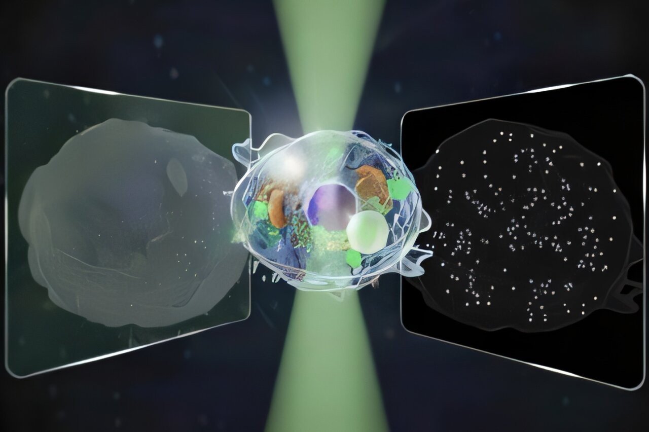
Researchers at the University of Tokyo have developed a novel microscope capable of simultaneously imaging structures across a 14-fold wider intensity range than conventional microscopes. Critically, this is achieved without the use of fluorescent dyes or other labeling agents, making it exceptionally gentle on living cells and ideal for long-term observation. The breakthrough, published in Nature Communications, addresses a fundamental limitation in modern microscopy: the trade-off between resolving large-scale cellular features and tracking individual nanoscale particles.
The Microscopy Dilemma
For centuries, microscopy has driven scientific progress. However, advanced techniques have historically required specialization. Quantitative phase microscopy (QPM) excels at imaging structures larger than 100 nanometers, providing a broad view of cells but lacking the sensitivity to detect smaller details. Interferometric scattering (iSCAT) microscopy, conversely, can track single proteins and nanoscale particles, but struggles to capture the comprehensive cellular context visible with QPM.
This divide forces researchers to choose between holistic snapshots and dynamic tracking – until now.
Bridging the Gap: Simultaneous Light Measurement
The research team, led by Kohki Horie, Keiichiro Toda, Takuma Nakamura, and Takuro Ideguchi, hypothesized that measuring both forward- and back-scattered light simultaneously could overcome this limitation. By analyzing how light interacts with a sample from both directions, they aimed to reveal a wide range of sizes and motions within a single image.
“I would like to understand dynamic processes inside living cells using noninvasive methods,” explains Horie, highlighting the motivation behind the work.
Validating the Microscope: Observing Cell Death
To test their new microscope, the team focused on a dynamic process: cell death. By recording a single image encoding information from both forward and backward-traveling light, they demonstrated the ability to quantify both the motion of larger cellular structures and the movements of tiny particles within the cell.
“Our biggest challenge,” Toda explains, “was cleanly separating two kinds of signals from a single image while keeping noise low and avoiding mixing between them.”
Quantifying Size and Motion
The resulting microscope not only captures the motion of structures across multiple scales but also estimates each particle’s size and refractive index. Refractive index, a measure of how light bends when passing through a substance, provides additional insight into the composition and properties of the observed particles.
This combined capability allows researchers to track dynamic changes within living cells without the artifacts introduced by fluorescent labeling. The unified approach promises to accelerate research in pharmaceuticals, biotechnology, and other fields requiring high-resolution, long-term cellular observation.
This development represents a significant step toward a truly versatile microscopy platform capable of bridging the gap between micro- and nanoscale imaging
